Published Papers by Dr Sommer
Published in the USA:
This clinical presentation represents a classic case of disc herniation with severe pain, numbness, and tingling, nights without sleep, inability to walk or sit and immense pain.
Surgery was the only recommendation, and the patient was placed on a waiting list at the Royal Melbourne Hospital, awaiting surgery.
The patient was referred to our office for treatment.
At the end of the treatment, the patient was asymptomatic, and a new MRI was ordered.
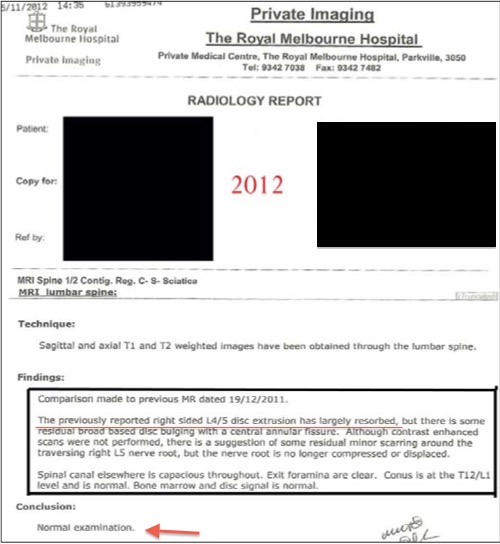
The new MRI was NORMAL. The previously reported large L4-5 disc was completely resorbed. (see picture below)


This case represents the growing approach of the conservative treatment of disc disorders with types of equipment like "DRS-9000", "Vax-D," "Flexion Distraction Decompression" and "Vertetrac." We are proud to have the latest equipment of the non-surgical decompression treatment of the discs.
Here is the link and the full case report including the MRI and CT:
http://www.coxtechnic.com/download-detail/disc-extrusion-resorbed-with-cox-technic-flexion-distraction-system-of-care
This clinical presentation represents a classic case of disc herniation with severe pain, numbness, and tingling, nights without sleep, inability to walk or sit and immense pain.
Surgery was the only recommendation, and the patient was placed on a waiting list at the Royal Melbourne Hospital, awaiting surgery.
The patient was referred to our office for treatment.
At the end of the treatment, the patient was asymptomatic, and a new MRI was ordered.
The new MRI was NORMAL. The previously reported large L4-5 disc was completely resorbed. (see picture below)
This case represents the growing approach of the conservative treatment of disc disorders with types of equipment like "DRS-9000", "Vax-D," "Flexion Distraction Decompression" and "Vertetrac." We are proud to have the latest equipment of the non-surgical decompression treatment of the discs.
Here is the link and the full case report including the MRI and CT:
http://www.coxtechnic.com/download-detail/disc-extrusion-resorbed-with-cox-technic-flexion-distraction-system-of-care
Another Publication
Published in the USA:
Spinal Canal Stenosis, Facet Arthropathy and Disc Prolapse Resulting in Foot Drop and Responding to Spinal Decompression
A 64-year-old white married female, presented with a complaint of left leg pain and weakness. The symptoms persisted for three weeks after gardening and lifting a heavy paving stone. The patient rated her symptoms on a visual analog scale as seven on a scale between one (no pain) and ten (worst pain). The patient reported not having these or similar symptoms in the past.
CT was ordered and shown Spinal Canal Stenosis at L4 5, and a left-sided disc prolapse with impingement of the left L4 root.
PHYSICAL EXAM:
SLR=60 on the left reduced reflex and foot drop
IMAGING


COMMENTS
The patient was treated with Non-Surgical Spinal Decompression and responded well to treatment. She was released asymptomatic at the end of treatment regaining complete strength in her foot.
Non-Surgical Spinal Decompression is based on the following:
1. Increases the intervertebral disc height to remove tension on the annular fibers and spinal nerve by increasing foraminal area and increasing circulation.
2. The intradiscal pressure within the nucleus pulposus drops from a positive of 25 mm Hg to a negative centripetal force within the nucleus pulposus to -192 mm Hg.
3. The area of the intervertebral foramen (osseoligamentous canal) increases up to 28%
4. Physiological range of motion is restored to the zygapophyseal joints via mobilization under distraction. This case represents the necessity of proper diagnostic evaluation even when the patient arrives from another physician.
http://www.coxtechnic.com/download-detail/case-report-91-spinal-stenosis-with-foot-drop-successfully-relieved-with-cox-technic
Spinal Canal Stenosis, Facet Arthropathy and Disc Prolapse Resulting in Foot Drop and Responding to Spinal Decompression
A 64-year-old white married female, presented with a complaint of left leg pain and weakness. The symptoms persisted for three weeks after gardening and lifting a heavy paving stone. The patient rated her symptoms on a visual analog scale as seven on a scale between one (no pain) and ten (worst pain). The patient reported not having these or similar symptoms in the past.
CT was ordered and shown Spinal Canal Stenosis at L4 5, and a left-sided disc prolapse with impingement of the left L4 root.
PHYSICAL EXAM:
SLR=60 on the left reduced reflex and foot drop
IMAGING
COMMENTS
The patient was treated with Non-Surgical Spinal Decompression and responded well to treatment. She was released asymptomatic at the end of treatment regaining complete strength in her foot.
Non-Surgical Spinal Decompression is based on the following:
1. Increases the intervertebral disc height to remove tension on the annular fibers and spinal nerve by increasing foraminal area and increasing circulation.
2. The intradiscal pressure within the nucleus pulposus drops from a positive of 25 mm Hg to a negative centripetal force within the nucleus pulposus to -192 mm Hg.
3. The area of the intervertebral foramen (osseoligamentous canal) increases up to 28%
4. Physiological range of motion is restored to the zygapophyseal joints via mobilization under distraction. This case represents the necessity of proper diagnostic evaluation even when the patient arrives from another physician.
http://www.coxtechnic.com/download-detail/case-report-91-spinal-stenosis-with-foot-drop-successfully-relieved-with-cox-technic